1. A Detailed Look Inside
Inside the Lungs
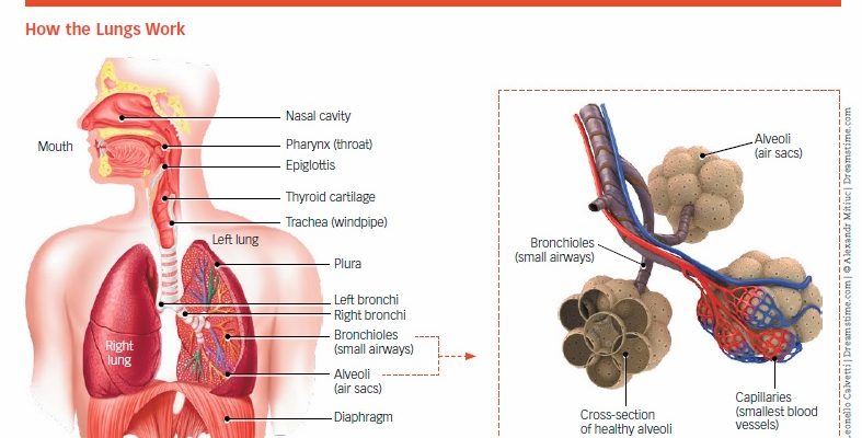
The lungs consist of two large, spongy organs divided into sections called lobes. The right lung has three lobes, and the left lung has two. Inside the lungs is a branching system of progressively smaller tubes (called bronchial tubes), which end in air sacs. Oxygen and carbon dioxide travel through these tubes (see Box 1-1, “How the Lungs Work”).
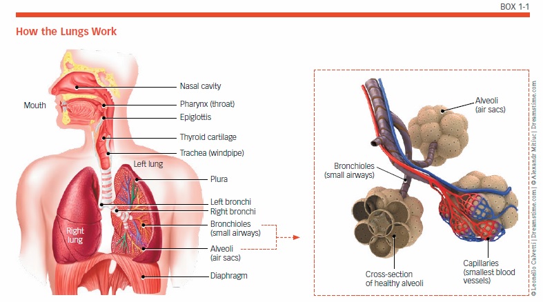
The system of bronchial tubes looks like the branches of a tree. The main trunk is the trachea, and it branches into two large tubes called the right and left mainstem bronchi, each of which leads into one of the two lungs. These bronchi branch off into smaller and smaller tubes called bronchioles. The trachea and the bronchial tubes (bronchi and bronchioles) are often referred to as “airways.”
When air (which contains oxygen) is breathed in through the mouth and nose, it passes through the voice box (larynx) and enters the windpipe (trachea), the opening of which is at the back of the throat. Air then travels down the trachea into the bronchial tubes in the lungs.
Oxygen In and Carbon Dioxide Out
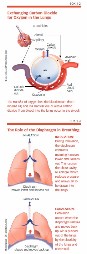
At the end of the bronchioles are clusters of tiny air sacs that look like bunches of grapes. These are called alveoli, and there are millions of them. When you inhale, these sacs expand as they fill with air. When you breathe out, the air sacs deflate. The alveoli have very thin walls, and are surrounded by capillaries (the smallest blood vessels). Where the alveoli and capillaries meet, gas exchange takes place (see Box 1-2, “Exchanging Carbon Dioxide for Oxygen in the Lungs”). Oxygen moves across the walls of the alveoli and enters the tiny capillaries, where it is absorbed into red blood cells. This “oxygenated” blood begins its journey to the heart, and then the rest of the body. At the same time, carbon dioxide from the blood in the capillaries passes into the alveoli. From there, it is expelled from the body through exhalation. This exchange of gases takes just a fraction of a second.
The Process of Breathing
Most people take 12 to 20 breaths every minute. This occurs automatically, but you can also consciously control your breathing, making it faster or slower and even holding your breath for a brief period. The automatic function of breathing is controlled by the brain stem (the lower part of the brain that connects to the spinal cord), along with other functions necessary for survival, including digestion, heart rate, and blood pressure. When the brain senses that there is too little oxygen or too much carbon dioxide, it sends signals to increase the speed and depth of breathing. Because active cells (including muscle cells) require more oxygen for energy and produce more carbon dioxide, breathing increases with exercise and slows down during rest.
The mechanical process of breathing depends on several muscles, the largest of which is the diaphragm. This dome-shaped muscle is positioned just below the lungs, and separates the chest cavity from the abdomen (see Box 1-3, “The Role of the Diaphragm in Breathing”). During inhalation, the diaphragm flattens out, causing the chest cavity to enlarge. This reduces pressure in the chest cavity and allows air to be drawn into the lungs. When the diaphragm relaxes, air is pushed out of the lungs. Other muscles also are at work during the process of breathing. When these muscles are working most efficiently, more air is pulled into lower lobes of the lung.
The Role of the Heart and Circulatory System
There are two types of blood vessels: Veins carry blood toward the heart, and arteries carry it away from the heart. Arteries bring blood that contains oxygen (oxygenated) to the tissues of the body, and veins carry blood that is void of oxygen (deoxygenated) and full of carbon dioxide back to the heart. Through the heart’s pumping action, blood that has been depleted of oxygen and contains carbon dioxide is sent from the heart into the lungs. Once gas exchange takes place in the lungs, the newly oxygenated blood returns to the heart. From there, the heart pumps the oxygenated blood into the aorta (the main artery) and out into the branching system of smaller and smaller arteries to reach the rest of the body (see Box 1-4, “Blood Flow: Heart and Lungs”).

Protective Mechanisms of Bronchial Tubes
Bronchi and bronchioles are essentially tubes with muscular walls. Lining the inside of these tubes are several layers of tissue. The innermost lining is called the mucosa, and it contains cells that help protect the lungs. One type of cell in the mucosa produces the sticky substance known as mucus. The mucosa also contains cells with hair-like projections called cilia. Mucus serves many useful functions. For example, it traps bacteria, pollen, and other particles that have been inhaled. These are then swept away from the lungs and up toward the mouth by the motion of the tiny cilia and cleared away, mostly through swallowing. Coughing can also get rid of mucus. When you get a cold or flu, excess mucus is created. The mucus (also called sputum) that comes up when you cough is helping to eliminate the infectious agent. Once the infection is gone, mucus production returns to its normal state and coughing eventually stops.
The bronchial tubes are surrounded by bands of muscle. Certain cells in the lining of the bronchial tubes have receptors that stimulate these muscles to contract and relax. For example, a type of receptor called a beta-adrenergic receptor causes muscles to relax, which widens the airway, making it easier to breathe. Another type of receptor, called a cholinergic receptor, causes the muscles to contract, narrowing the airway and making it harder to breathe. Normally, this keeps irritants out of the lungs. For people with obstructive lung diseases, this narrowing can make it even more difficult to breathe.
The post 1. A Detailed Look Inside appeared first on University Health News.
Read Original Article: 1. A Detailed Look Inside »
Powered by WPeMatico